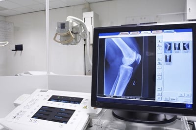
X-rays have become widely used in nearly every medical field, including oral health. X-rays are not the actual picture that is produced, but rather are the rays sent through the portion of the body requiring internal study. While they are not visible to the naked human eye, using specific technology, a picture (radiograph) can be created in shades of white, grey, and black. Here is a look at understanding dental X-rays.
What is the purpose of dental X-rays?
Dental X-rays can be used in several different ways. One of their main uses is during recommended annual checkups. These scans allows the X-rays to flow through the softer tissues of the mouth (the gums) and record images of the teeth. In this way, potential issues can be noticed and managed before they become a real problem.
While extraoral radiographs are taken outside the mouth, intraoral radiographs are taken from inside the mouth and provide a more detailed image of the oral structures. Dental radiographs are largely intended as preventative measures, but may be used for diagnosis as well.
What kinds of dental X-rays are there?
Depending on the reason a patient is visiting the dentist, different types of X-rays may be necessary. Here are some of the most common ones.
- Bite-Wing: Bite-wing radiographs are one of the most common type of dental X-rays. Images are taken of the back teeth, which allows dentists to see any decay that might be forming in the there, as well as getting a look at the way the back teeth touch each other during biting. Generally four of these are required to get a full view.
- Panorex: Panorex is an extraoral imaging technique. Rather than taking an image from the inside, an X-ray “camera” moves around the outside of the mouth taking a panoramic view. Different issues can be seen using panorex radiographs as opposed to full set radiographs, particularly issues like impacted teeth. Panoramic radiographs also capture the sinus and nasal areas, and jaw. These images are often useful for orthodontia or other implant surgeries.
- Full Set: A “full set” X-ray utilizes 14 to 20 different radiographs to get a full view of all the teeth, as well as the under- and overlying bone. It is often used as an initial tool for diagnosis of many different issues, from cavities to tumors.
- Periapical: A periapical radiograph is used when there is a problem with a particular tooth. Periapical X-rays provide a detailed image of the one tooth, from the bone underneath to the crown, or external tip of the tooth.
- Occlusal: Occlusal radiographs examine the base, or floor, of the mouth. This is useful for seeing how tooth development is progressing in children.