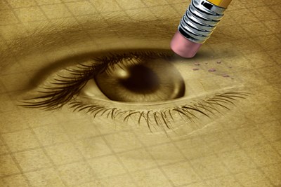
Blindness is a heartbreaking disability, whether it’s congenital or obtained later in life. But that doesn’t mean you shouldn’t understand it. Here are ten terms to help you understand blindness.
- Cataracts: Cataracts affect more than half the world’s population, with more than 24 million Americans over the age of 40 having been affected by them. The lens of the eye that is normally clear becomes cloudy over the course of several years. It slowly becomes more and more difficult to see faces, drive, or read. Cataract surgery has become extremely safe and effective, however, by removing the cloudy lens and replacing it with a new, artificial lens that allows clear sight again.
- Glaucoma: Glaucoma is another leading cause of blindness and vision loss worldwide. Rather than starting in the eye, glaucoma is a group of disorders that occur from damage to the optic nerve which sends the eye’s information to the brain. There is no cure for glaucoma, although treatments can help slow the progression of the disease. Unfortunately, even with treatment 10-15% of people will still completely lose vision in at least one eye.
- Optic Nerve: Being blind can mean there’s something awry in multiple places. There may be an issue with the eyes, or with a part of the brain that processes vision. The optic nerve is one of the cranial nerves that connects the eyes to the brain. When it becomes damaged (such as during glaucoma), what the eyes see can’t be sent to the brain, interfering with vision.
- Retina: The retina is one of the layers of the eyeball responsible for vision. It resides near the back and is largely made up of rods and cones (photosensitive cells that decipher light and send signals up to the optic nerve). These signals are sent to the occipital lobe and visual cortex in the brain, allowing us to “see.” The retina can become torn or detached from age or injury, interfering with clear vision.
- Macula: Also called the “fovea centralis,” the macula is a small spot in the retina that allows you to see fine details in your central vision. There are multiple types of macular degeneration, a disorder in which the macula begins to essentially disintegrate. It can destroy the central vision of the eye, making it nearly impossible to recognize faces, read fine print, or engage in activities that require small detail (like cross stitching). The peripheral vision, however, often stays intact, as it is controlled by a different area of the eye.
- Cornea: The clear dome on the outside of the eyeball is the cornea. It is made up of five layers, each with a different job. It is part of the protective anatomy of the eye, and its shape helps focus light into the retina.
- Corneal Opacity: Corneal opacity is a clouding of the cornea, generally as a result of injury or infection. This cloudiness inhibits the passage of light to the retina, making vision blurry or impossible. Corneal opacities may result from particles in the eye scratching the surface, chemicals irritating the cornea, intense light refracting off reflective surfaces (snow blindness), as well as infections like ocular herpes or conjunctivitis when left untreated.
- Retinitis Pigmentosa (RP): Retinitis pigmentosa are genetic disorders that cause the retina to effectively disintegrate. The rods and cones slowly die out, leading to vision loss. New advances in stem cell research, prosthetic eyes, and gene therapy have begun to find potential cures for blindness as a result of different types of RP.
- Visual Acuity Test: When diagnosing some eye disorders, an ophthalmologist will see how a patient performs on a visual acuity test. These are used at annual eye exams as well. It consists of a chart containing a series of letters that grow smaller on each line. The point at which the patient can no longer read the letters helps eye doctors see how strong their vision is -- and how much it has declined since their previous visit.
- Slit Lamp Examination: A slit lamp examination is another optic diagnostic tool. It is a specialized magnifier with a strong light attached. This allows the eye doctor to examine the structures at the front of the eye by lighting up and magnifying the cornea, iris, and lense. Slit lamp examination makes it much easier to see abnormalities on the surface of the eye, such as cataracts.