Since the accidental invention of X-rays by a German physicist, a whole variety of useful places have been found for the technology. Despite the frequency of their use, X-rays are not well understood. Here is a look at 10 terms to help you learn more about medical X-rays.
- Wavelength: Each type of light ray has a specific length between the highest points of its “wave.” The light we can see has a relatively long wavelength. However, the shorter wavelength of X-rays allow them to pass through low density, nonmetallic objects—like skin and organs, allowing healthcare professionals to take pictures of internal structures.
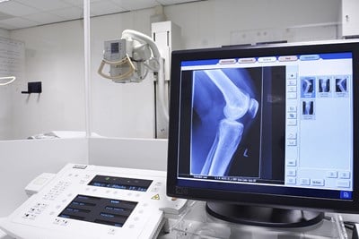
- Source: In order to actually take these pictures, the X-rays must come from somewhere. An X-ray source can be several things, but it generally consists of an X-ray generator. X-ray generators contain a vacuum tube which uses a cathode and anode to collect and direct electrons. The X-rays are carried by photons to the area being examined, and a negative (black and white) image is created.
- Detector: Once the X-ray source emits the X-rays, there has to be something to receive them. Because of the differing density of bodily structures, the X-ray waves pass through them differently. The detector measures each newly arrived ray, which is then displayed in black and white on the radiograph.
- Radiological density: Radiological density is the differing “thickness” of bodily structures. Depending on how dense each part is, and what element it’s made of, the radiological density differs. The more radiologically dense matter is, the whiter it appears on the final picture. For example, because bones are mostly calcium, they are much more radiologically dense than skin. X-rays pass through organs, but not bone, thus bone shows up white and organs much darker.
- Radiograph: A radiograph is what most people think of when they hear the term "X-ray." A single moment in time is captured when the X-rays pass from source to detector through the body. The black, white, and grey image is created, and healthcare professionals can view bone fractures, tumors, and other issues.
- Computed tomography: Computed tomography (or CT) scans utilize X-rays, as well as computers, to create a different sort of image from the standard black and white snapshot. Images are taken horizontally to create a much clearer and more detailed image of the body.
- Fluoroscopy: Fluoroscopy is a type of X-ray that uses injected or orally administered contrast dye to create real-time images in tandem with CT scans. Instead of a single shot of internal structures, fluoroscopies, taken against a fluorescent screen. They can be used for diagnosing issues or for medical procedures, like angioplasy, in which it is necessary to see what’s going on inside while doing the procedure.
- Radiation therapy: While many X-ray technologies are used for diagnosis, they can also be used for some treatment options. The ionizing radiation X-rays produce can be targeted at cancerous cells or tumors. The radiation then damages the cancer’s DNA, hopefully destroying it in the process.
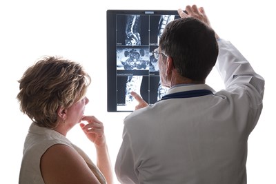
- Radiographer: Radiographers are health care professionals, but not doctors, who receive specific training in utilizing imaging techniques. Although they are fully qualified to create the images required, diagnosis and interpretation are not part of their role in the medical field.
- Radiologist: Radiologists are doctors who are specially trained to diagnose conditions through imaging techniques, such as X-rays or CT scans. They receive unique training concerning the effects of radiation on the body and know how to create and understand the many different sorts of imaging tests.