X-rays were an inadvertent discovery by a German physicist. While working with cathode rays, he stumbled upon the unknown, unseen light waves he dubbed “X”-rays. X-rays, as it turned out, have much shorter wavelengths than waves that are visible to the human eye. They can penetrate low-density, nonmetallic matter—like skin, but not bone.
While X-rays are used in several diagnostic tests, there are also treatments available through X-ray technology. Here is a look at some of the more common X-ray uses.
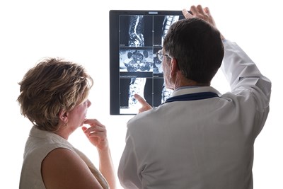
Medical X-Rays
X-rays can be used in several diagnostic situations. Radiographs are taken by emitting X-rays from a source, through the body part requiring an internal image, and onto a detector. The different density of the body structures are displayed by differing shades. of black and white For example, bones appear white because of the dense calcium they contain—contrasting sharply with the darker areas the X-rays pass through easily.
In this way, it is possible to see damage to internal structures: fractures and breaks, foreign objects, tumors, pneumonia, and other injuries.
Mammograms are radiographs specially intended for the discovery and diagnosis of breast cancer. If a tumor is present, the contrast will be sharper than surrounding tissue. Microcalcifications, or tiny flecks of calcium which indicate particular types of cancer, can also be seen on mammograms.
Combined Technologies
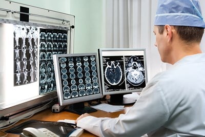
X-rays are often used in conjunction with other types of diagnostic tools. Combining forces like this allows doctors to form a more detailed, accurate view of what is wrong with the patient.
Computed Tomography (CT) scans combine radiographs with computers to provide different views of the body. Rather than a 2D image, the computer processing allows professionals to “generate a series of cross-sectional images of the body that can later be combined to form a three-dimensional x-ray image,” according to the National Institute of Health (NIH). In this way, doctors have more angles to view what’s going on inside the body. Pictures are taken horizontally, and provide a much more detailed view of any body part.
While radiographs are a single snapshot of the body at the moment of capture, fluoroscopies utilize a contrast solution to make a “movie” instead of a photograph. A solution is swallowed or injected intravenously, and the X-rays are sent continuously against a fluorescent screen to allow real time viewing of internal structures. Even the blood’s movement can be viewed this way.
Radiation Therapy
X-rays produce ionizing radiation, which means X-ray technology can also be used to treat cancer. X-ray radiation can be injected directly into the blood, or waves can be aimed at a tumor or area where the cancer is located. The large amount of radiation damages cancer DNA, hopefully destroying it in the process. Radiation therapy may be used alone or in tandem with additional types of cancer treatment.