There are many sorts of imaging tests available for medical diagnostics. And while most people who think “ultrasound” think “pregnancy,” there are many uses for ultrasounds. There are also many ways to perform an ultrasound, as they come in different types. There are many benefits for choosing ultrasounds (or sonograms) over other types of imaging technology.
How does an ultrasound work?
While x-rays make pictures of bones, sometimes you need to be able to see more than that. Enter ultrasounds. Extremely high frequency sound waves are sent into the body, bounce of bones or organs, and send echoes back to the machine to create a 3D image or video of what’s going on with the soft tissue inside the body. It can tell not only what it looks like and how big it is, but also the consistency of the organ in question.
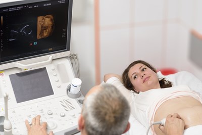
When you go in to get an ultrasound, the sonographer (an ultrasound technician) will smear the area being recorded with jelly. This allows the transducer that sends out the sound waves to move smoothly across the skin. Most ultrasound sessions take less than an hour, including cleaning off the jelly.
What can ultrasounds see?
Nearly any organ can be viewed by ultrasound. According to the Radiological Society of North America, the list of body parts that an ultrasound can be an effective tool for is quite long, but far from comprehensive. Their list includes the “heart…, blood vessels, liver, gallbladder, spleen, pancreas, kidneys, bladder, uterus, ovaries, unborn children…, eyes, thyroid and parathyroid glands, scrotum,” and the brains and hips of infants.
Ultrasounds are also useful during biopsies and for diagnosing many heart conditions. Additionally, it is possible to view “blockages to blood flow (such as clots), narrowing of vessels, tumors and congenital vascular malformations,” and areas where blood is abnormal.
What types of ultrasound are there?
In some instances, it is necessary to get closer to the organ in question to get a clear picture for optimal viewing. As such, there are multiple ways of attaching the transducer to probes which are inserted into the body.
- Transvaginal ultrasound: The probe is inserted into the vagina to improve images of the reproductive system.
- Transrectal ultrasound: The probe is inserted into the rectum for prostate diagnostics.
- Transesophageal ultrasound: The probe is inserted down the esophagus to get closer to the heart.
- Doppler ultrasound: Rather than the use of a probe, blood flow throughout the body’s vessels are assessed.
What are the benefits of an ultrasound?
The Radiological Society of North America states that there are “no known harmful effects on humans” from an ultrasound. It doesn’t require the use of any needles (although there are the probes to consider), and there is no radiation involved as with some other imaging technologies. This is not only better for the person of whom the ultrasound being taken but especially for a sonogram’s use during pregnancy with an unborn baby. There is very little pain associated with it, although there may be some discomfort depending on the exact procedure. It allows a clear view of things x-rays are unable to pick up, and the image being viewed is in “real time,” meaning what you see on the screen is exactly what’s going on in your body at that very moment.