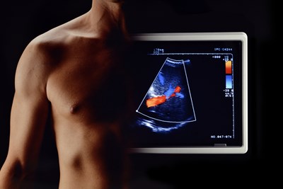
An ultrasound is a noninvasive imaging technology that uses high-pitched sound waves to take real-time images and videos of organs and other tissue inside the body. There are a variety of imaging techniques, but, according to the Radiological Society of North America, ultrasounds (also called sonograms) provide a variety of medical services with “no known harmful effects on humans.” They can be used diagnostically, evaluatively, and to assist with internal medical procedures.
How an Ultrasound Works
Depending on what part of the body is being sonogrammed, that area is covered in jelly. This not only allows the transducer to slide across the skin without friction, but also facilitates sound waves. The transducer or probe is a microphone-shaped piece of equipment that sends out sound waves at a frequency so high humans can’t hear it. The waves bounce off to be picked back up by the transducer, and the echoes are then translated into a picture or video on a monitor of what’s occurring internally at the exact moment of the ultrasound.
Diagnostics and Evaluation
The Radiological Society of North America lists the following nonexclusive collection of internal body parts that ultrasounds are especially useful for examining:
- Heart and blood vessels
- Liver
- Gallbladder
- Spleen
- Pancreas
- Kidneys
- Bladder
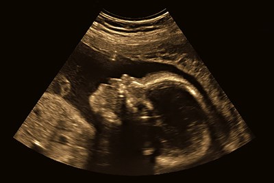
- Uterus, ovaries, and fetuses
- Eyes
- Thyroid and parathyroid glands
- Scrotum
Ultrasounds may be necessary for doctors to examine a patient’s organs and insides for a variety of reasons. Some illnesses can cause damage to certain organs. There may be internal problems that can’t been seen or otherwise examined. Ultrasounds can also allow physicians to rule out certain conditions or disorders if visible symptoms are not present. Some body parts require certain types of probes in order to get closer to the organ in questions.
Medical Assistance and Treatment
Because of the nature of ultrasounds, videos and pictures are in “real time.” This means that if the sonographer notices an abnormal internal mass, it can be easier to take a biopsy of the mass while using ultrasound technology. A biopsy involves taking a small sample of the abnormal tissue to test it for problems. Ultrasounds are often great options for those in need of internal medical care. While the benefits in most situations far outweigh the risks, it is still best to discuss your options with your doctor to make the right choice.