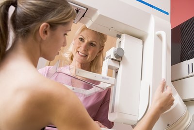
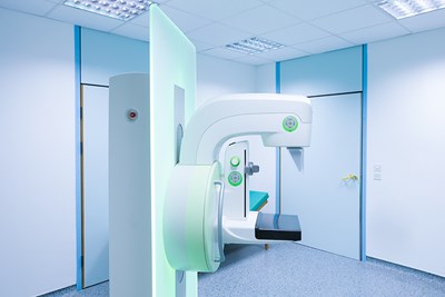
Mammograms are X-ray images of your breasts that are used to detect tumors or other abnormalities that may indicate cancer. There are different types of mammograms that are used for different purposes.
Screening Mammogram
Screening mammograms typically involves taking two X-ray pictures of each breast to look for any signs that point to breast cancer. Women with larger breasts may need more than two pictures in order to include as much breast tissue as possible. The goal of screening mammograms is to detect cancer before there are any clinically detectable signs or symptoms. Finding breast cancer when it is still in the early stages makes it much easier to treat and prevent.
During a mammogram screening exam, the radiologist is looking for anything that could be evidence of cancer that requires further testing to be sure. Possible findings of screening mammograms include:
- Calcifications: These are benign calcium deposits that develop in the ducts or other tissues of the breast.
- Masses: These are lumps that may or may not be cancerous.
- Distorted tissues: This result could indicate a tumor that has invaded the neighboring tissues.
- Dense areas: If a specific area appears denser than the rest of the breast, this may be cause for concern, especially if it has developed since your last mammogram.
Any of these results would require further testing in order to determine if the abnormalities are cancerous or benign. The next step would likely be a recommendation for a diagnostic mammogram.
Diagnostic Mammogram
Diagnostic mammograms involve taking as many X-ray images as necessary in order to get a good view of an area that is concerning your doctor. If you have an abnormal area that is found during a screening mammogram, you will typically need a diagnostic mammogram. Additionally, women who are experiencing any abnormal issues with their breasts such as a lump or discharge from the nipple would likely be recommended for a diagnostic mammogram as well.
During some diagnostic mammograms, special images such as spot views or magnification views are used to focus on a smaller area of concern and make it easier to evaluate. It is also likely that you will have to undergo other tests as well, such as an ultrasound or MRI (magnetic resonance imaging).
The standard interpretations of diagnostic mammograms include:
- It could reveal that upon closer examination, an area that looked abnormal during regular mammogram screening is actually perfectly normal. If this is the case, you will simply go back to your normal routine of yearly screening.
- It could reveal that the area of concern is most likely not cancer. However, the radiologist will likely wants to watch the area closely just to be sure, so you will probably be asked to come in for another mammogram in about four to six months.
- It could suggest that a biopsy is needed in order to definitively determine whether the abnormal area is cancerous. However, you should keep in mind that just because your doctor recommends a biopsy, this does not mean that you have cancer.