A pulmonary embolism (PE) occurs when a clot travels from one of the larger blood vessels (generally in the deep veins of the legs) and lodges in the smaller pulmonary vessels of the lungs. A pulmonary embolism can be fatal. While there are a variety of treatments, the efficacy of those treatments derives in part from a swift and accurate diagnosis. Unfortunately, it can be difficult to diagnose PE, particularly if you have other cardiovascular issues. Here’s a look at how a pulmonary embolism is diagnosed.
Understanding the Symptoms
While unusual symptoms are your key to realizing something isn’t right, giving your doctor a clear and accurate depiction of what your body is undergoing can go a long way toward a diagnosis. Recognizing the signs of PE can help your doctor understand where to start looking and what tests you should undergo. The most common symptoms associated with a pulmonary embolism include a cough that produces blood, chest pain (much like the pain that occurs during a heart attack), and shortness of breath.
According to the Mayo Clinic, less common, but nonetheless important, symptoms include leg pain, cyanosis, fever, perspiration, dizziness, and heart palpitations. Make a note of any symptoms you experience so that your doctor has a solid picture from which to make a diagnosis.
Running Blood Tests
Blood testings can be used to detect clotting disorders, such as Factor V Leiden, that place individuals at an increased risk of developing the condition. The presence of clots can change the way the body “answers” to blood tests. Treating PE focuses largely on thinning the blood to prevent new clots from forming. While some cases may make use of thrombolytics (clot dissolvers), the body can actually dissolve blood clots on its own. When the substance in charge of dissolution, D-dimer, is very high, it can be indicative of a serious blood clot—although this is not the only reason D-dimer levels may rise.
Other blood tests may be used to monitor oxygen and carbon dioxide levels in the blood. PE interferes with the way your lungs send out freshly oxygenated blood to the rest of the body and, potentially, the way it gets rid of carbon dioxide waste. Too little oxygen or too much carbon dioxide may be indicative of a pulmonary embolism.
Using Imaging Tests
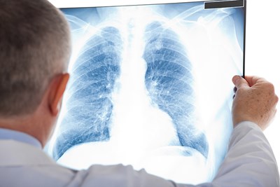
While blood tests and physical exams can go a long way towards helping your doctor identify a proper diagnosis, imaging tests can help truly narrow down concerns. An x-ray of the chest is not necessarily helpful for diagnosis, but it is helpful for ruling out other issues with similar symptoms. Duplex ultrasonography is often used to diagnose deep vein thrombosis (DVT), a common contributing factor of PE. A spiral CT scan, with or without contrast, may be given to get a much closer look at the blood vessels than a standard CT scan offers. The helical scan takes images in a spiral, allowing greater precision, according to the Mayo Clinic. An MRI may be used on pregnant women or others to whom contrast or radiation may pose additional risks.
A pulmonary angiogram is generally considered to provide the clearest view of the lungs and their blood vessels, but it also requires someone very skilled in the technique to administer it properly. It also comes with a handful of serious risks. Among these risks is a change in the way your heart beats or a reaction to the dye in the face of pre-existing kidney problems. As such, most doctors opt for other, easier imaging techniques before resorting to a pulmonary angiography for diagnostic purposes. An angiogram involves the use of a catheter (a long, flexible tube) which is threaded through one of the larger veins in the body—typically the groin. Once the catheter has followed the line of vessels into the pulmonary region, a contrast dye is injected. As the dye spreads, X-rays are taken to watch the way it spreads and to find out if or where the clots are located.