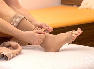
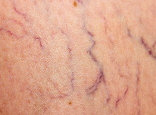
When the one-way valves in our veins weaken due to old age or excess pressure, this causes blood to begin to flow backwards and accumulate in pools. This, in turn, leads to a condition known as varicose veins. Diagnosing this condition is straightforward and fairly simple, although a doctor may recommend additional tests to determine the extent of a patient’s varicose veins.
Physical Examination
Because varicose veins are normally so visibly prominent, many cases can be diagnosed by a physical exam alone. Doctors will normally examine patients while they have their legs in a variety of positions, such as sitting, standing, and dangling. Doctors will often ask questions about any symptoms, such as pain or swelling.
Duplex Ultrasound
After the initial physical exam, many physicians will recommend a duplex ultrasound, which can monitor the flow of blood in a patient’s legs and detect blood clots that can be the result of varicose veins. With a duplex ultrasound, a patient’s blood vessels are mapped via traditional ultrasounding techniques, while a Doppler ultrasound is then used to monitor the flow of blood within the veins. Like a pregnancy ultrasound, doctors run a device over the affected area—typically the legs.
Angiogram
If a doctor is having difficulty getting the full picture of a patient’s condition with a duplex ultrasound, he or she can recommend further testing—namely, an angiogram. During this procedure, dye is injected into the veins and then documented via X-ray imaging. This is used to definitively determine whether a patient is suffering from varicose veins or another related condition, such as spider veins. Since this procedure is so invasive, it’s only rarely employed.